Overview
What Is Fluorescence Lifetime?
Fluorescence lifetime (τ) is the average time a fluorophore remains in its excited state before emitting a photon. Unlike fluorescence intensity, fluorescence lifetime is independent of fluorophore concentration and excitation power. Instead, it directly reflects conformational changes and the local microenvironment. Fluorescence lifetime enables discrimination of the fractional contributions of different lifetime components and sensitive detection of subtle lifetime changes, providing a stable and specific quantitative readout for in vivo studies.
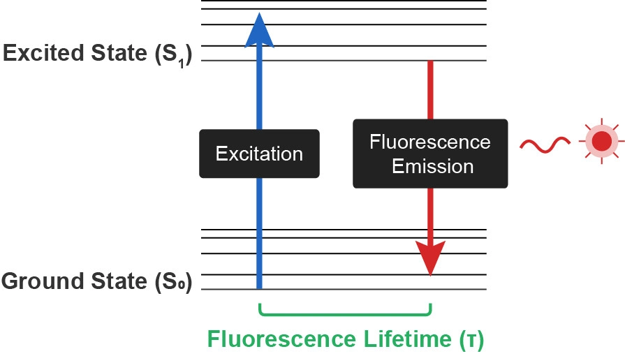
Fig.1 Principle of fluorescence lifetime. Fluorescence lifetime (τ) is the average time a fluorophore remains in its excited state before emitting a photon.
Why Fluorescence Lifetime?
Unlike conventional fluorescence intensity, fluorescence lifetime is an intrinsic property of the fluorophore — independent of probe expression, photobleaching, and motion artifacts. This enables:
-
Quantitative, cross-sample comparable measurements — without normalization to baseline
-
Stable long-term recordings — ideal for sleep, chronic disease models, and longitudinal studies
-
Sensitive detection of dynamic changes in brain analyte levels via lifetime-based probes
Two Mainstream Measurement Methods
Time-Domain
Time-domain methods excite the sample with a pulsed light source and directly record the temporal profile of fluorescence decay. The most widely used implementation is time-correlated single photon counting (TCSPC): the time difference between each emitted photon and the preceding excitation pulse is measured, a decay histogram is constructed, and lifetime values are extracted by exponential fitting. Time-domain methods offer high temporal resolution, time individual photons, typically provide superior signal-to-noise ratio under low photon flux, and resolve multiple lifetime components in multiexponential decays with high precision.
Frequency-Domain
Frequency-domain methods excite the sample with a sinusoidally modulated continuous light source and derive fluorescence lifetime from the phase delay and modulation depth of the emission relative to the excitation. Frequency-domain systems are relatively simple, offer fast acquisition, and do not require costly pulsed lasers, making them particularly suitable for high-speed wide-field imaging of samples with high photon flux.
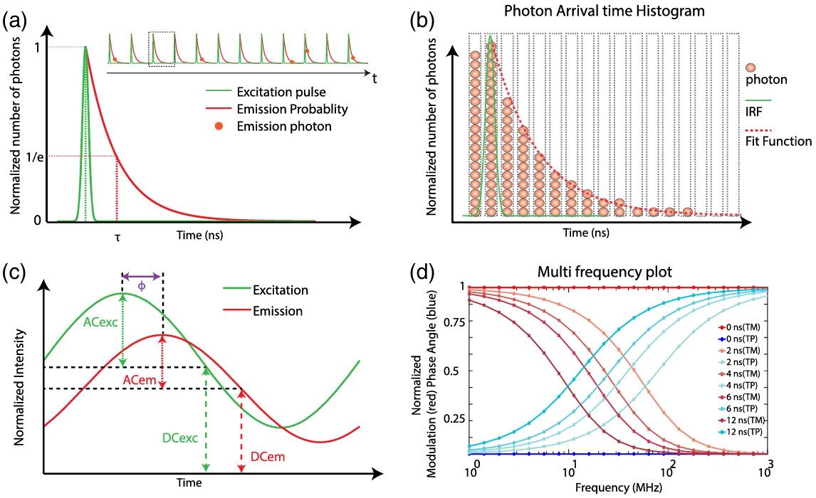
Fig.2 Schematic of time-domain (TCSPC) and frequency-domain FLIM[1]. (a) TCSPC: pulsed excitation and fluorescence decay, lifetime (τ) is defined at 1/e. (b) Photon arrival time histogram with IRF (green) and fit function (dotted red). (c) Frequency-domain: sinusoidally modulated excitation and phase-shifted emission. (d) Modulation and phase versus frequency for different lifetimes.
How to Choose Between the Two Methods
Time-domain (TCSPC) offers higher temporal resolution, times individual photons, provides superior signal-to-noise ratio for weakly fluorescent samples, and resolves multiple lifetime components with high precision.
Frequency-domain offers faster acquisition, performs well with samples of high photon flux, and can be implemented without pulsed lasers.
|
Comparison
|
Time-Domain (TCSPC)
|
Frequency-Domain
|
|
Lifetime measurement accuracy
|
High
|
Good
|
|
SNR for weakly fluorescent samples
|
Excellent
|
Relatively limited
|
|
Resolution of multiple lifetime components
|
High precision
|
Requires multi-frequency modulation or phasor analysis
|
|
Acquisition speed
|
Relatively fast
|
Fast
|
|
High photon flux samples
|
Good
|
Excellent
|
|
Laser requirement
|
Pulsed laser
|
Modulated continuous light source
|
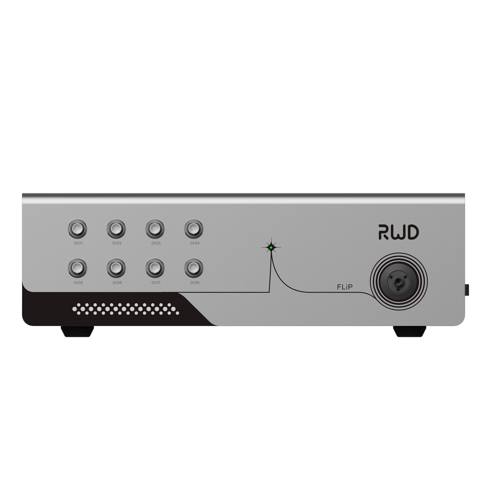
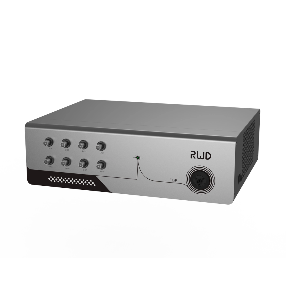
The RWD Fluorescence Lifetime Photometry System employs the industry-recognized gold standard, TCSPC, to directly detect fluorescence lifetime as a readout of neural activity, supporting long-term, stable recordings in freely moving mice. The system measures fluorescence lifetimes in the 1–10 ns range and is compatible with the vast majority of fluorescence lifetime probes available on the market, providing a stable and reliable quantitative solution for in vivo neuroscience research.
Highlights of Fluorescence Lifetime Photometry System
 |
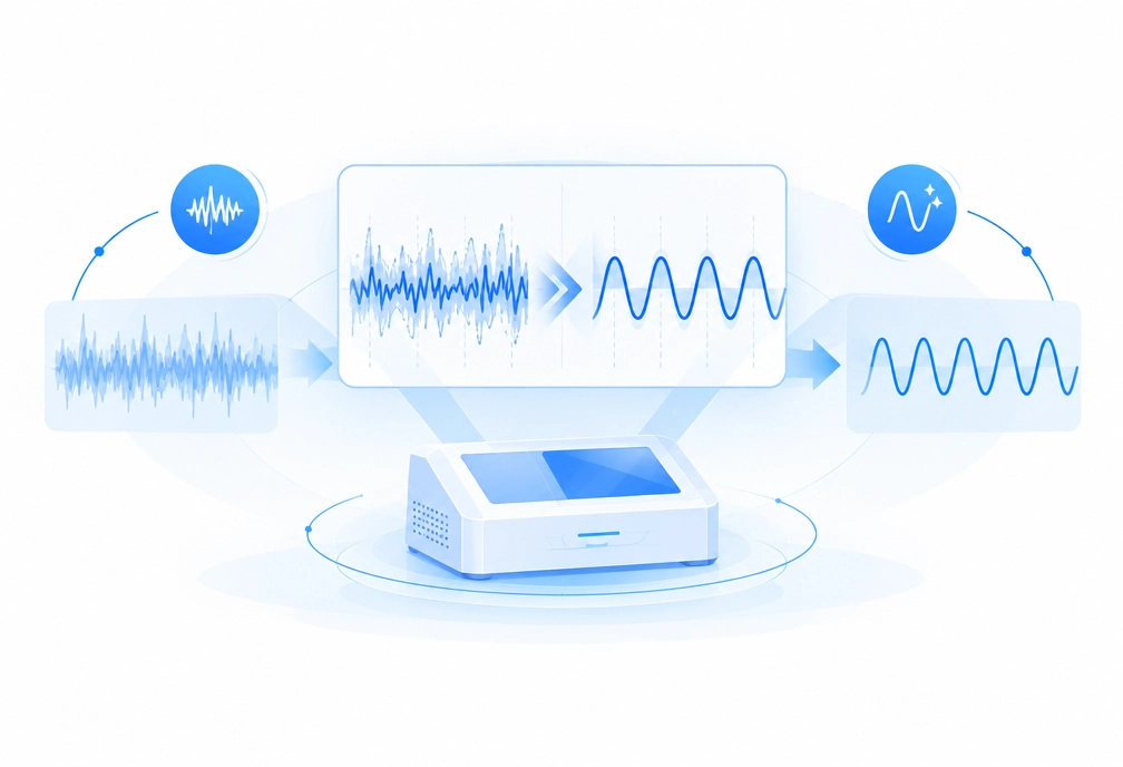
Exceptional System Stability
Consistent system performance minimizes the impact of device-state fluctuations, supporting reliable comparisons across longitudinal recordings.
|
Ultra-Low System Noise
Low system noise helps distinguish true signals from background interference, preserving data integrity even under weak-signal conditions.
|
 |
 |
Reliable Long-Term Recording
Stable core components help minimize baseline drift over consecutive weeks, supporting dependable long-term data collection.
|
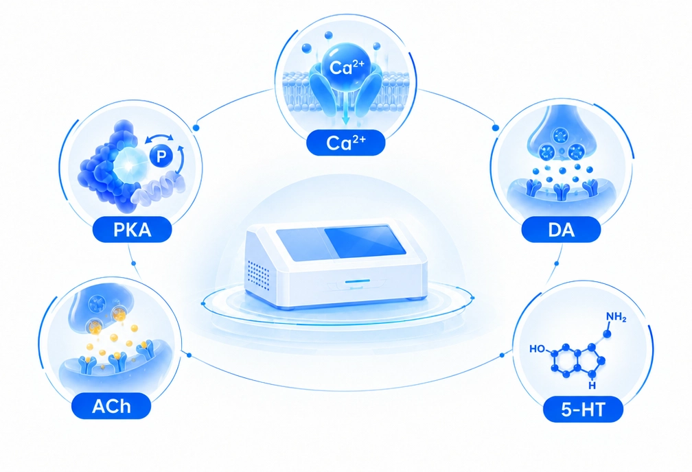
Broad Biosensor Compatibility
Supports the fluorescence lifetime design range of mainstream biosensors, enabling diverse detection applications with a single system.
|
 |
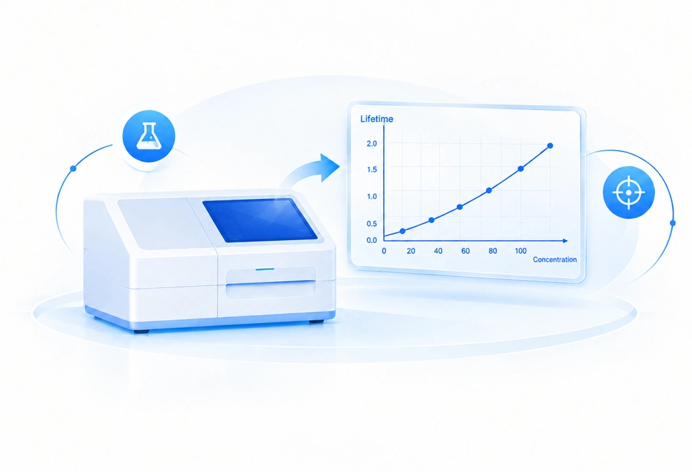 |
Direct Quantitative Analysis
The planned standard curve function will enable direct concentration readouts for more straightforward and precise quantitative analysis.
|
Application Examples
Fluorescence lifetime photometry (FLiP) enables stable, quantitative, and cross-sample comparable in vivo measurements of neural signals in freely moving animals. The following three examples illustrate its applications across different research directions.
(1) Neuromodulator Measurements Across Animals and Chronic Time Scales [2]
Researchers at Washington University School of Medicine systematically evaluated the application of fluorescence lifetime measurements to neuromodulator sensors. They found that multiple intensity-based sensors (e.g., GRAB ACh3.0, GRAB 5-HT2h, GRAB NE2m, GRAB DA2m) also exhibit fluorescence lifetime responses, with GRAB ACh3.0 showing the largest lifetime dynamic range.
Using FLiP, the authors measured GRAB ACh3.0 lifetime and intensity in the hippocampal CA1 region of freely moving mice. Unlike intensity, fluorescence lifetime remained stable across fluctuations in laser power, differences in sensor expression levels, and changes in expression over weeks. After pooling data across conditions, only lifetime significantly distinguished behavioral states such as running versus resting and NREM versus REM sleep. This study demonstrates that fluorescence lifetime is a reliable readout for comparing neuromodulator levels across animals and chronic time scales.
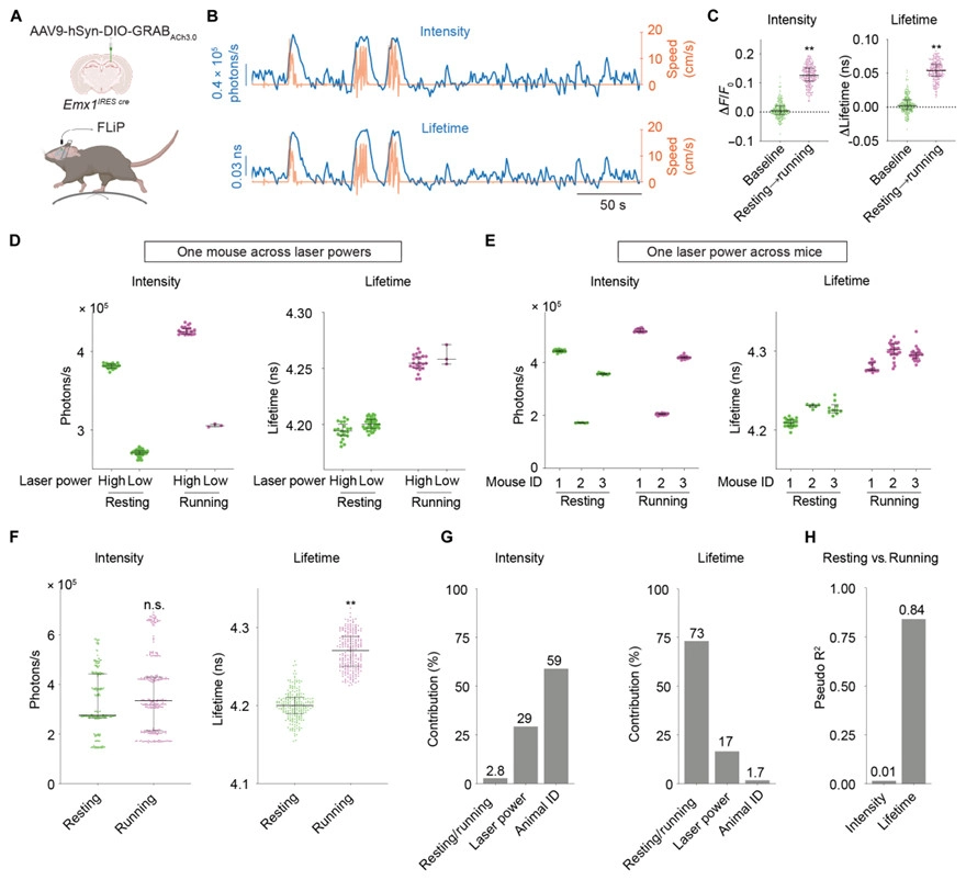
Fig. 3 Fluorescence lifetime of GRAB ACh3.0 correlates with running versus resting states accurately despite varying laser powers and varying sensor expression levels across mice in vivo[2]
(2) Revealing Abnormal Striatal PKA Activity in Akap11 Mutant Mice, a Genetic Model of Psychiatric Disorders [3]
Researchers at the Broad Institute and collaborating institutions used FLiP to measure PKA activity dynamics in the striatum of Akap11 mutant mice, a genetic model carrying loss-of-function mutations in a risk gene for schizophrenia and bipolar disorder.
The authors expressed the PKA activity reporter FLIM-AKAR in the nucleus accumbens and performed FLiP measurements in freely moving mice. Results showed that PKA activity was persistently elevated and stable across weeks in Akap11 mutant mice, whereas the PKA-insensitive mutant FLIM-AKAR T391A showed no differences across genotypes. Following dopamine receptor antagonist administration, D2 antagonists further enhanced the already elevated PKA activity in mutant mice, while D1 antagonists produced more pronounced inhibition of PKA activity in mutants. Behaviorally, mutant mice exhibited abnormally prolonged locomotor responses to amphetamine. This study reveals that Akap11 deficiency leads to abnormal striatal dopamine and PKA signaling, providing molecular mechanistic evidence for the dopamine hypothesis of psychiatric disorders.
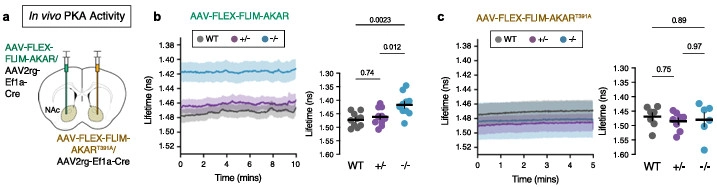
Fig. 4 Fluorescence lifetime photometry (FLiP) in Akap11 mutant mice reveals constitutively elevated PKA activity and functional alterations to striatal circuitry[3]
(3) A Novel Calcium Lifetime Sensor LifeCamp Enables Absolute Measurement of Transient and Baseline Calcium Signals [4]
Researchers at Harvard Medical School developed LifeCamp, a novel green high-speed calcium lifetime sensor, and validated its performance in freely moving mice.
Using FLIPR, the authors measured LifeCamp lifetime in ventral tegmental area (VTA) dopamine neurons and in D1R- and D2R-expressing spiny projection neurons of the nucleus accumbens. They found that baseline calcium levels in VTA dopamine neurons were significantly higher than in both types of nucleus accumbens neurons. In addition, LifeCamp detected calcium transients evoked by food pellet consumption and foot shocks, and revealed slow fluctuations in baseline calcium during repeated feeding. This study demonstrates that LifeCamp enables absolute measurement of both baseline and transient calcium changes across multiple in vivo conditions, providing a new tool for studying fast and slow dynamics of neuronal activity.
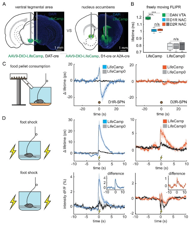
Fig. 5 LifeCamp measured in the VTA and NAC of freely moving mice[4]
Together, these three examples demonstrate the value of fluorescence lifetime photometry in cross-animal and cross-time-scale consistency validation, mechanistic studies of psychiatric risk genes, and the development and application of novel lifetime sensors, highlighting its unique advantages for resolving in vivo neural signals and opening new possibilities for the field.
Fluorescence Lifetime Photometry System Applications

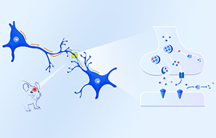
Calcium or Neurotransmitter Signal Detection
Compatible with calcium and neurotransmitter signal detection, including mainstream probes such as jGCaMP and dLight3.8, and next-generation fluorescence lifetime probes in development. |

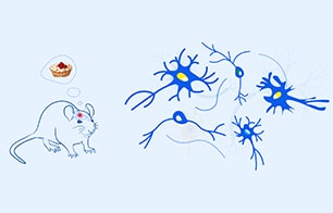
Neural Circuit Research
Applicable to reward, punishment, emotion, and social behavior paradigms, including long-term recordings such as sleep studies, with signals unaffected by photobleaching. |


Investigation of Pathological Mechanisms of Neurological Disorders
Enables stable cross-sample comparisons independent of probe expression, facilitating direct comparisons between disease models and normal controls. |

Development of Novel Fluorescent Probes
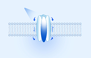
Supports fluorescence lifetime probe development and validation, including probe screening, characterization, and methodology establishment. |

In Vivo Quantification of Signaling Molecules in the Brain
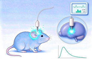
Converts measured fluorescence lifetime into substrate concentration, advancing analysis from relative fluorescence intensity changes toward absolute quantification. |
[1] Datta R, Heaster TM, Sharick JT, Gillette AA, Skala MC. Fluorescence lifetime imaging microscopy: fundamentals and advances in instrumentation, analysis, and applications. J Biomed Opt. 2020;25(7):1-43. doi:10.1117/1.JBO.25.7.071203
[2] Ma P, Chen P, Tilden EI, Aggarwal S, Oldenborg A, Chen Y. Fast and slow: Recording neuromodulator dynamics across both transient and chronic time scales. Sci Adv. 2024;10(8):eadi0643. doi:10.1126/sciadv.adi0643
[3] Song BJ, Ge Y, Nicolella A, et al. Elevated synaptic PKA activity and abnormal striatal dopamine signaling in Akap11 mutant mice, a genetic model of schizophrenia and bipolar disorder. Nat Commun. 2025;16(1):10793. Published 2025 Nov 28. doi:10.1038/s41467-025-66504-2
[4] Bart Lodder, Michelle Raghubardayal, Sanika Ganesh, Xintong Cai, Josh Stern, Michael Sherman, Paul Rosen, Tarun Kamath, Isa Hartman, Michelle Siegel, Joshua Timmins, Roger Adan, Mark Andermann, Bernardo L. Sabatini. bioRxiv 2025.12.23.696288; doi: https://doi.org/10.64898/2025.12.23.696288