In 1665, Robert Hooke, a British scientist, discovered and named the cells. Subsequently, German biologists Matthias Jakob Schleiden and Theodor Schwann proposed the cell theory, giving everyone a clearer understanding of the cell, a relatively independent unit that makes up our lives.
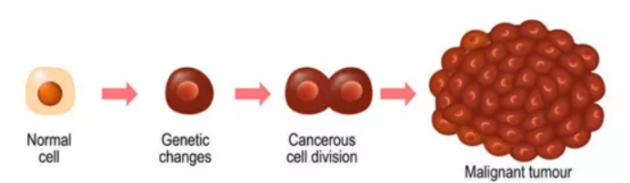
In order to better understand the essence of life and reveal the laws of life activities of cells, scientists have carried out a series of researches on cell proliferation, movement, metabolism, death and other activities. In fact, as early as the 19th century, some people proposed the concept of separating living cells from tissues, but the culture of animal cells was not studied until 1950. From simple observation of cell development, to isolation of primary cells from tissue, to obtaining continuous cell lines that can be passaged continuously. The transformation of cells from an illusory concept to an indispensable experimental material for in vitro research experiments has run through the entire history of biological development. Cells used for scientific research are mainly divided into two types: primary cells directly isolated from tissues and purchased cell lines.
Usually, the cells from the first to tenth passages isolated from the tissue are collectively referred to as primary cells, and most of the primary cells will gradually stagnate, senesce and die after this, and a very small number of cells that can survive to the fiftieth generation. After fifty generations, the genetic information of cells has changed, and they have become a continuous cell line. It has the characteristics of immortality, and some cells have lost contact inhibition, can grow in multiple layers, and can even cause tumors after allogeneic inoculation.
Continuous cell lines have always been the first choice for cell-level studies due to their immortality and rapid division. In addition, it has the characteristics of convenient culture, wide variety, fast growth rate, low cost, and rapid research, which makes various continuous cell lines widely used. For example, Hela cells, as the first human cell line that can be passaged indefinitely, help scientists to more efficiently focus on human cell diseases and the mechanism of action of active drugs, and are widely used in oncology, immunology, virology and other disciplines.
In recent years, studies have found that although continuous cell lines are full of treasures, there are still some limitations in their application.
Limitation 1: The biology of the cell is altered
During the research process, a large number of samples are often required, so the rapid propagation and expansion of continuous cell lines are required. And the continuous passaging process is prone to mutations, unrestricted passage may lead to changes in the genotype and phenotype of the cell line, thereby affecting the experimental results. At the same time, the biological characteristics of immortalized cell lines and living cells are quite different, and they cannot fully represent the real environment in animals. Primary cells are considered to be more representative of the in vivo tissue situation and have been legally recognized in some countries/regions (Human Tissue Act 2004, UK), especially in the early toxicity assessment tests of drugs, compared to cell lines, using primary cells isolated directly from patient tissue is more appropriate.
Limitation 2: cell line cross-contamination
When it comes to cell contamination, everyone’s first reaction is that pathogenic microorganisms cause the failure of cell experiments. However, what is mentioned here is not to say that the bacterial proliferation in the culture system affects the experiment, but the cross-contamination between cell lines, that is, the cell lines used for the experiment may be mixed with other types of cells. This problem may be caused when the cell line is established, or it may be caused by some improper operations, such as co-culture of multiple cells at the same time, reuse of consumables, or caused by related cell culture products. Every inadvertent operation can lead to the failure of the entire experiment.
In 2011, the United States issued a national standard for cell STR identification. Although many researchers are still unaware of the seriousness of the problem, relevant institutions have begun to sound the alarm:
In both the December 2014 and February 2015 issues of the journal Science, articles were published explaining the seriousness of cell cross-contamination and misidentification, and highlighting the consequences of contamination. It not only wastes time and energy, but also the research results cannot be reproduced, and its impact is very serious. Therefore, authoritative institutions such as NIH and ATCC have repeatedly issued calls for researchers to identify cells. In April 2015, Nature announced that all its journals would require authors to identify the cell lines used in their papers. Then in June, it was reported that a scientist withdrew the Nature paper due to cell line problems. In October 2017, an article published in the journal PLoS ONE stated that there was cross-contamination of the HeLa cell lines used in more than 30,000 papers, rendering the research results invalid[1]. Another study in 2019 showed that at least 24% of human cell lines were contaminated with HeLa[2].
Limitation 3: Cell Application
Many cells fail to proliferate in vitro, and each experiment requires isolation of primary cells from fresh tissue. Such as neurons, skeletal muscle cells, cardiomyocytes, pericytes, and terminally differentiated hepatocytes, etc.
In addition, the state of primary cells is closest to the physiological environment in the body and can maintain certain tissue biological characteristics. Therefore, it is the best choice for evaluating drug efficacy and virulence, and at the same time, the probability of cross-contamination is greatly reduced. In addition, for studies related to oncology and immunology, if living animal models are involved, the isolation of primary cells is also required. However, the difficulty of culturing primary cells is much higher than that of ordinary cell lines. Meanwhile, the isolation and preparation of primary cells has always been an urgent problem to be solved. Under the premise of ensuring reproducibility, how to efficiently prepare a single-cell suspension with high activity?
RWD single cell suspension dissociator easily solves this problem. The self-developed tissue processing tube, enzymatic digestion kit and built-in optimization programs for preparing high-activity single-cell suspension and tissue homogenate.
The instrument can quickly complete the preparation of single-cell suspension with high activity and good uniformity, increasing the repeatability of experiments. It has four independent channels, and the heating jacket can improve the efficiency of tissue processing. It is widely used in immunology, oncology, neurobiology and other research fields.
After obtaining the single cell suspension, the cells need to be counted and analyzed for viability and placed in a culture system or corresponding buffer system. RWD automatic cell counter C100can accurately count primary cells. With multiple fluorescence channels, it can quickly quantitatively analyze cells in suspension, and simultaneously display brightfield and fluorescence images, clearly showing the counting results and cell morphology. It is very suitable for immunology, vaccine development, cell therapy, tumor research, stem cell and metabolic research and other fields.
Cell culture is the last step in the primary cell separation process.RWD CO2 incubatorcan precisely control the temperature and carbon dioxide concentration, and can maintain a high-humidity environment. The HEPA high-efficiency filter can effectively remove particulate pollution in the air, and the 140°C dry heat sterilization can realize regular sterilization and maintenance of the incubator, providing a stable and clean culture environment for primary cell samples.
【References】
1. Serge P. J. M. Horbach, Willem Halffman. The ghosts of HeLa:How cell line misidentification contaminates the scientific literature. PLoS ONE, Published:October 12, 2017, doi:10.1371/journal.pone.0186281
2. Lin J, Chen L,Jiang W, Zhang H, Shi Y, Cai W. Rapid detection of low-level HeLa cell contamination in cell culture using nested PCR. J Cell Mol Med.2019;23(1):227–236. doi:10.1111/jcmm.13923
1.Fill in the form and our experts will get back to you ASAP!
2.Ask about An Equipment
3.Wondering which equipment to conduct your researches or perform your experiments? Our
sales reps will try their best to share their knowledge with you!
4.Get Technical Support
5.An RWD equipment is not performing? Talk to our support team to get instant feedback!